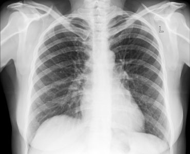


Angiography, also known as arteriography, is the examination of the flow of blood through veins and arteries with the help of a special form of X-ray. This is used for investigating kidneys, heart, legs as well as brain very closely. This is done by injecting into the bloodstream, a dye called contrast medium, and then X-rays are taken.
Any abnormal condition is displayed during angiography. It is also used to find out the diseases that can cause the blood vessels to appear differently. Angiography can also be used to check the presence of any coronary heart disease.
Angiography helps to identify the presence of thrombosis that is caused by aneurysm. It can also help to detect if any abnormal developments like cysts, tumours or congenital defects or any injury to the organ is present.
Angiography helps to investigate
Cerebral bleeding
The condition of the arteries which supply blood to the brain
If any abnormal arteries are present around brain.
Necessity
Angiography may be used to detect if there is any blockage in the arteries or if there is narrowing of the arteries. Since the arteries are the ones supplying blood to all the parts of human body, the health of the arteries is very important.
Any abnormality in the arteries is harmful for our health.
Any severe problem with the arteries can affect the limbs, heart, internal organs like kidney and liver as well the brain. Some other conditions due to malfunctioning of arteries are heart attacks, strokes, gangrene and organ failure.
How is angiography done
For a child, angiography is usually done under the general anaesthesia. But in most of the cases, adults as well as older children are given local anaesthesia for performing angiography.
A needle is inserted into the artery in the leg, groin or arm. Then a thin, long guide wire, which guides to the blood vessel needing examination, is inserted. A catheter is pushed over the wire to get it into the right position. After that wire is taken out and the contrast medium is allowed to flow into the blood vessels thought the catheter.
The image is viewed on the screen and video film containing the data is made which is used for future investigations.
Another method is the use of Digital-subtraction angiography which produces the detailed blood vessel image by discarding the unwanted details from the surroundings.
Another form used by the ophthalmologists is fluorescein angiography which is used in the examination of the micro blood vessels that are present in the iris, chord and retina of our eyes. This method is very helpful for investigating the condition called posterior vitreous detachment.
Usage
Uses are
- Renal angiography
- Coronary angiography
- Pulmonary angiography
- Extremity angiography
- Cerebral angiography
- Lymphangiography
- Left heart ventriculography
- Right heart ventriculography
- Aortography
Arteriography can be used
- For detecting aneurysms,
- For looking at pancreas and liver
- For detecting tumours
- Locating internal bleeding sites
- locate the site of internal bleeding,
- to help if surgery is needed.